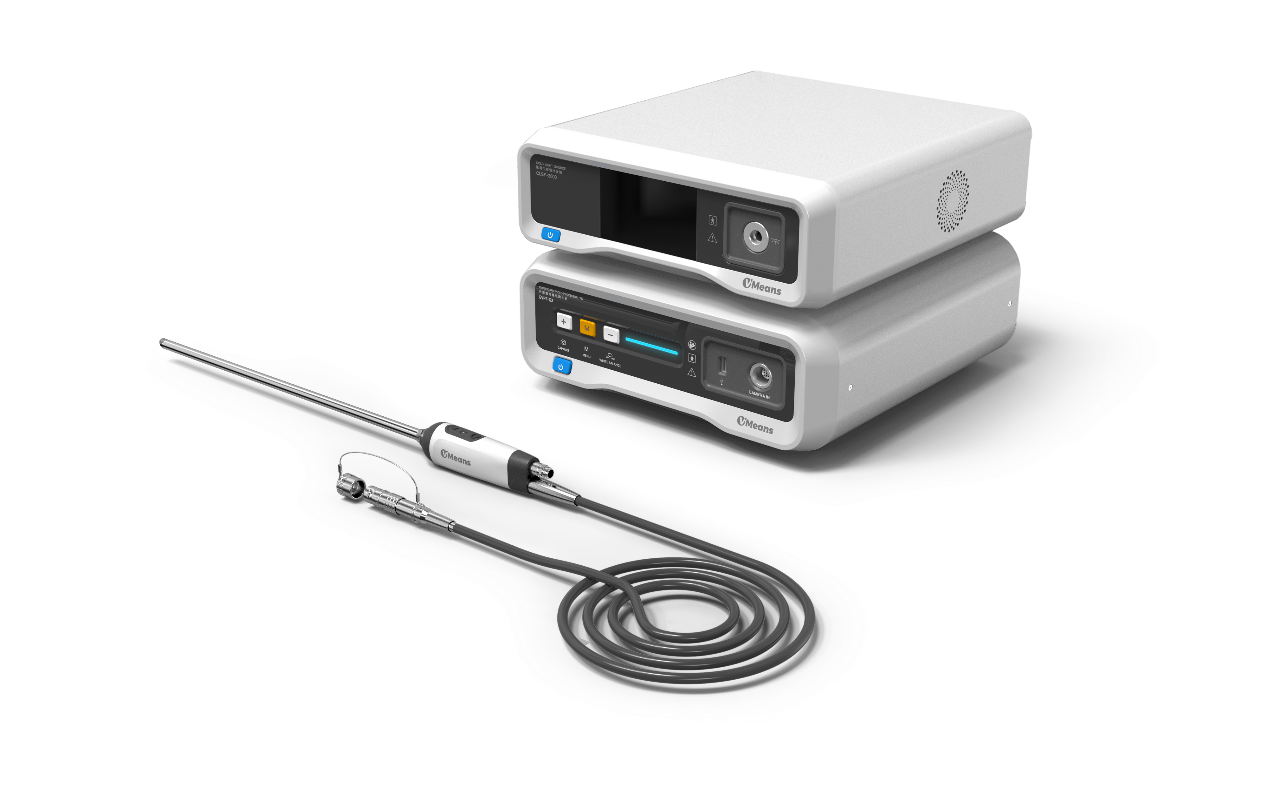
On July 24th, in Shanghai, China, the real-time fusion fluorescence 3D electronic laparoscopic endoscope imaging system, developed by Shanghai MicroPort MedBot (Group) Co., Ltd. (hereinafter referred to as "MicroPort® MedBot®") for use with the Toumai® laparoscopic surgical robot, was officially approved for market release. This image processing system is compatible with the medical electronic laparoscope developed by MicroPort® MedBot® and the fluorescent contrast agent Indocyanine Green (ICG).

▲The Graph of Real-time Fusion Fluorescence 3D Electronic Laparoscopic Imaging System
ICG is a water-soluble, near-infrared fluorescent contrast agent, approved for disease diagnosis by the US FDA in 1959. In an aqueous solution, it naturally decomposes into colorless derivatives and binds with proteins in the blood and tissues within the human body. When stimulated by near-infrared light with wavelengths of 600-900nm, ICG emits fluorescence with a penetration depth of 1-5 cm in tissues. It is metabolized relatively quickly in normal human cells with a half-life of 3-4 minutes. In solid tumors, it exhibits both fluorescence and high permeability and retention effects, which hold significant clinical value. Especially in the process of metastatic lymph node tracing, tumor metastasis diagnosis, and pelvic lymph node dissection of prostate cancer, ICG can provide clear lymphatic drainage pathways and help identify the relationships between important structures such as lymph nodes and blood vessels, ensuring that metastatic lymph nodes are not missed. In procedures such as radical prostatectomy, radical cystectomy, radical gastric cancer resection, and others, it assists clinical surgeons in identifying the tumor lymphatic system, observing tissue blood perfusion, and precisely locating tumor boundaries in real-time, which avoids positive resection margins, lowers the surgery risk, makes radical cancer surgery more thorough with more evident clinical effects.
The laparoscopic surgical robots currently available on the market, equipped with fluorescent endoscopes, use black-and-white mode-switching fluorescence technology. This technology, through image sensor configuration, transfers the near-infrared spectrum to the visible spectrum channel, thereby obtaining fluorescence information while losing color details. Consequently, it is impossible to observe color images and detailed tissue structures, making it difficult for surgeons to operate directly under the fluorescence image. Surgeons must frequently switch between fluorescence and white light images to confirm tissue dissection and the degree of positive margins, which increases the complexity of the operation and prolongs the surgery time.


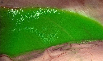
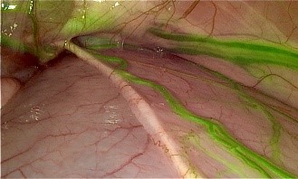
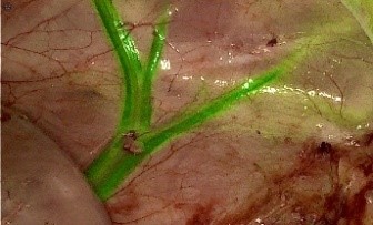
▲The Graph of Real-time Fusion Fluorescence Imaging
As a new-generation fluorescence technology, real-time fusion fluorescence technology independently captures images using both visible and near-infrared spectra. By combining multispectral fusion principles with innovative fluorescence algorithms, this technology seamlessly integrates visible light images with fluorescence images, providing real-time synchronized focal plane response and consistent focal plane display. This allows surgeons to observe both fluorescence images and clear non-fluorescent tissue details within a single frame, overcoming the barrier of being unable to perform surgery under fluorescence imaging. However, achieving both clear fluorescence boundaries and high fluorescence sensitivity in fluorescence imaging surgery is often challenging. After years of foundational research, the MicroPort® MedBot® team has surpassed extreme filtering performance at the 10-6 level, achieving the high fluorescence sensitivity and a stable signal range. This enables precise capture of clearly defined low-dose micro-metastatic lesions throughout the entire depth of field. Compared to black-and-white mode-switching fluorescence technology, real-time fusion fluorescence allows for tissue recognition, lesion identification, lesion removal, and residue confirmation all within a single frame, eliminating the need for intraoperative switching and significantly enhancing surgical convenience.
The approval of the real-time fusion fluorescence 3D electronic laparoscopic endoscope imaging system developed by MicroPort® MedBot® marks a significant advancement in China’s surgical robots through "independent innovation." This technology not only enhances the intelligence and safety of surgical robots but also exemplifies the exploration of China’s surgical robots with cutting-edge technology and innovative surgical models. It aims to provide more advanced technological services to a wide range of surgeons, benefit a larger patient population, and promote the advancement of surgical medical services.
-
 2024-04-03MicroPort® MedBot® Releases 2023 Annual Performance
 -
 2023-09-28MicroPort®MedBot™ (02252.HK) Included in Hang Seng Artificial Intelligence Theme Index
 -
 2023-08-01Headline Report on the People’ Daily – MicroPort® MedBot® Sends Out Loudest Voice of Building China into a Strong Cyber Power and Benefiting People with 5G Remote Surgery


 Hu ICP Bei No. 20013662 HGWA Bei No. 31011502015178
 " are registered trademarks of Shanghai MicroPort Medical (Group) Co., Ltd.” . They have been authorized to be used by Shanghai Microport Medbot (Group) Co., Ltd., and no other party shall use such trademarks without prior written permission thereof.
